IISER Pune · BRIC-NCCS Pune
How membranes hold their shape — and when they let go.
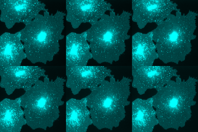
Biological membranes define the shape and boundaries of cells and organelles. Driven by the hydrophobic effect, lipids self-assemble into fluid bilayers without need of a template. This fluidity gives the membrane a selective permeability barrier — it blocks the free passage of ions and solutes while resisting mechanical stresses such as bending and vesiculation. These biophysical properties make lipid bilayers remarkably robust, which is likely why evolution settled on them as the enclosure for all living cells.
Yet for all their stability as barriers, membranes are constantly and deliberately challenged in the course of normal physiology. At the nanoscale, transient changes in permeability allow the controlled leak of ions and solutes — essential for setting membrane potential in excitable cells and organelles. At the microscale, the bilayer must bend and deform substantially during the budding and scission events that drive vesicular transport.
Specialized proteins manage both kinds of disruption. The Pucadyil Lab works to identify these molecular factors and to understand how they function and are regulated. We map these pathways through biochemical screens that pinpoint candidate proteins, in vitro reconstitution to probe their mechanism, and studies in yeast and mammalian cells to establish their physiological roles.
From the lab
Recent news
Meghadeepa and Sannidhya show that Dynamin-2 acts as a fission factor for SNX1-coated endosomal tubules — out now on bioRxiv.
Collaborative work with the Srivastava lab shows how phosphoinositide protonation states dictate AKT1 binding with the membrane, with Meghadeepa as lead experimentalist — out now on bioRxiv.
Shilpa and Uma show that yeast dynamin Vps1 drives the membrane constriction and fission required for endosomal protein sorting, published in EMBO Journal.
Thomas takes over as Director, BRIC-NCCS.
Collaborative work with the McPhee lab shows that division-arrest induced filamentation protects uropathogenic E. coli from killing by the cathelicidin antimicrobial peptide LL-37, with Meghadeepa among the contributing authors — out now on bioRxiv.
Keerti's work on membrane remodeling by the Bacterial Dynamin-like Protein (BDLP) from Nostoc punctiforme is out as a preprint on bioRxiv.
Meghadeepa's study on the division of labor among fission dynamins based on substrate size is published in Biochemistry.
Collaborative work with the Nandicoori lab shows that Wag31, a membrane tether, is crucial for lipid homeostasis in mycobacteria, published in eLife.
Collaborative work identifying how acute diacylglycerol production activates membrane-shaping proteins that drive mitochondrial tubulation and fission, published in Nature Communications.
What we study
Identifying membrane‑integrity‑disrupting (MID) proteins
Our central objective is to find the molecular factors that deliberately disrupt membrane integrity — and to understand what that disruption does for the cell. We build an in vitro platform that behaves like a real membrane, then watch it change shape in real time.
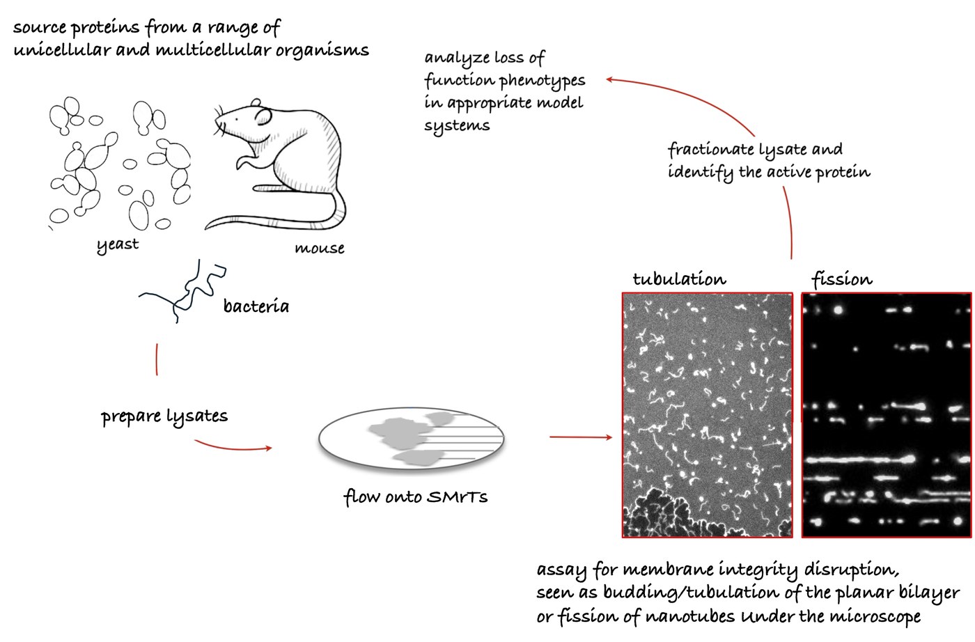
Platform — Supported Membrane Templates (SMrTs) are an adaptable in vitro system that generates everything from flat bilayers to highly curved nanotubes, so a candidate protein's effect on membrane shape can be watched directly under the microscope.
Workflow — Cell and tissue lysates are screened on SMrTs; morphological change is captured by real-time fluorescence microscopy, the responsible protein is pinned down by biochemical fractionation and mass spectrometry, then its physiological role is tested in budding yeast and mammalian cells.
Tubulation and fission
Two membrane-remodeling events recur across our projects — tubulation and fission.

Credit - Soumya Bhattacharyya
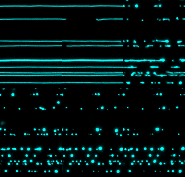
Credit - Srishti Dar
Support
Funding
75 and counting
Publications
Reverse chronological, from preprints back to 2004. Search by author, title, or journal.
Who we are
Team
Graduate students
Lab staff
Alumni
MS graduates
In the classroom
Teaching
Cell Biology
Membrane structure, transport, intracellular compartments, protein sorting, and vesicular traffic, through to cell-cycle dynamics, division, communication, junctions and adhesion, cytoskeleton regulation, motor proteins, cell migration, and an introduction to mechanical forces in cellular systems.
- Alberts et al., Molecular Biology of the Cell (2007 ed.)
- Watson et al. (2007 ed.)
- Lodish et al. (2007 ed.)
- plus course-specific literature reviews
Advanced Biochemistry
Two halves: membrane biochemistry — physical principles of membrane formation, protein insertion, lipid–protein organization, and reconstitution — and metabolism, spanning amino acid, lipid, carbohydrate, nucleotide and glycogen pathways, glycolysis, the citric acid cycle, and oxidative phosphorylation.
- Alberts et al., Molecular Biology of the Cell
- Mouritsen, Life — As a Matter of Fat
- Yeagle, membrane structure
- Metzler, Biochemistry, plus contemporary research articles
Lab-internal
Resources
Working inventories and shared documents for current lab members. Most links live on Google Drive and are access-restricted — ask a lab member for the current link if yours doesn't work.
Inventories
Data & protocols
- Shared data folders
- Protocols document
- Analysis macros
- SAM7 (iiserpune.in)
- PhD trajectory tracker
Prospective members: ask about access during your visit or interview.
Find us
Contact
BRIC-National Centre for Cell Science
Savitribai Phule Pune University Campus,Ganeshkhind Road, Pune 411007,
Maharashtra, India
IISER Pune
Dr. Homi Bhabha Road, Pashan,Pune 411008,
Maharashtra, India